
Digestive Health
Digestive disorders can have a significant impact on a horse’s health, comfort and overall well-being. Signs can sometimes be subtle, ranging from changes in appetite or body condition to recurrent colic, behavioural changes or reduced performance.
At ESMP, digestive investigations are tailored to each horse and may combine different diagnostic techniques to better understand the underlying cause of the problem.
What is gastroscopy?
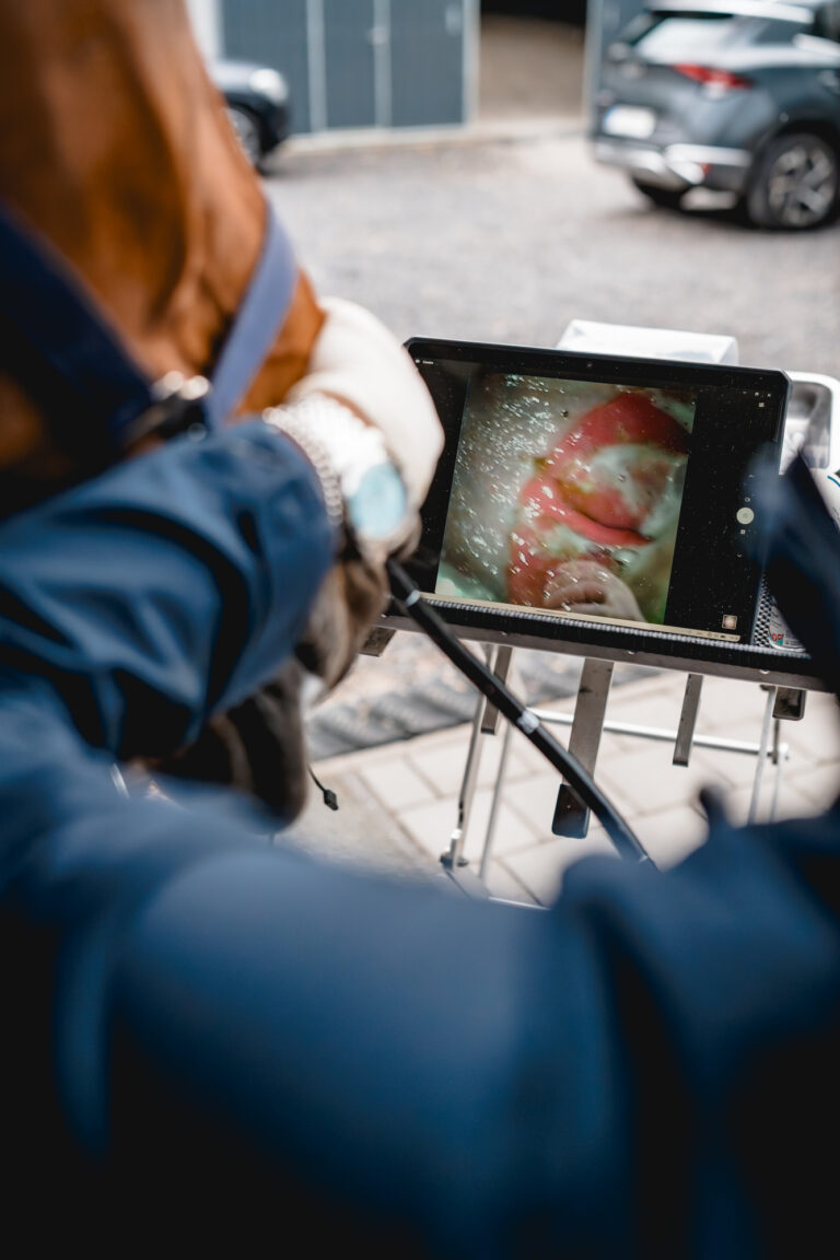
Gastroscopy is an endoscopic examination that allows direct visualisation of the upper digestive tract, including the oesophagus and stomach.
It is particularly useful for identifying abnormalities of the gastric mucosa, including Equine Gastric Ulcer Syndrome (EGUS), as well as assessing the oesophagus when an abnormality or previous obstruction is suspected.
Gastric disease is common in horses, particularly when factors such as feeding practices, training, travel, competition and management influence their natural digestive physiology. Because clinical signs can be subtle or non-specific, gastroscopy remains the most reliable way to determine whether gastric lesions are present and to assess their location and severity.
When is gastroscopy recommended?
Gastroscopy may be recommended in horses presenting with:
- Reduced or variable appetite
- Unexplained weight loss or poor body condition
- Recurrent or mild colic
- Behavioural changes or signs of discomfort
- Reduced willingness to exercise
- Unexplained poor performance
- Suspected or previously diagnosed gastric disease
The findings allow treatment and management recommendations to be tailored to the individual horse.
How is a gastroscopy performed?
For the stomach to be examined properly, it must be empty. The horse therefore needs to be fasted before the examination according to the instructions provided by our veterinary team.
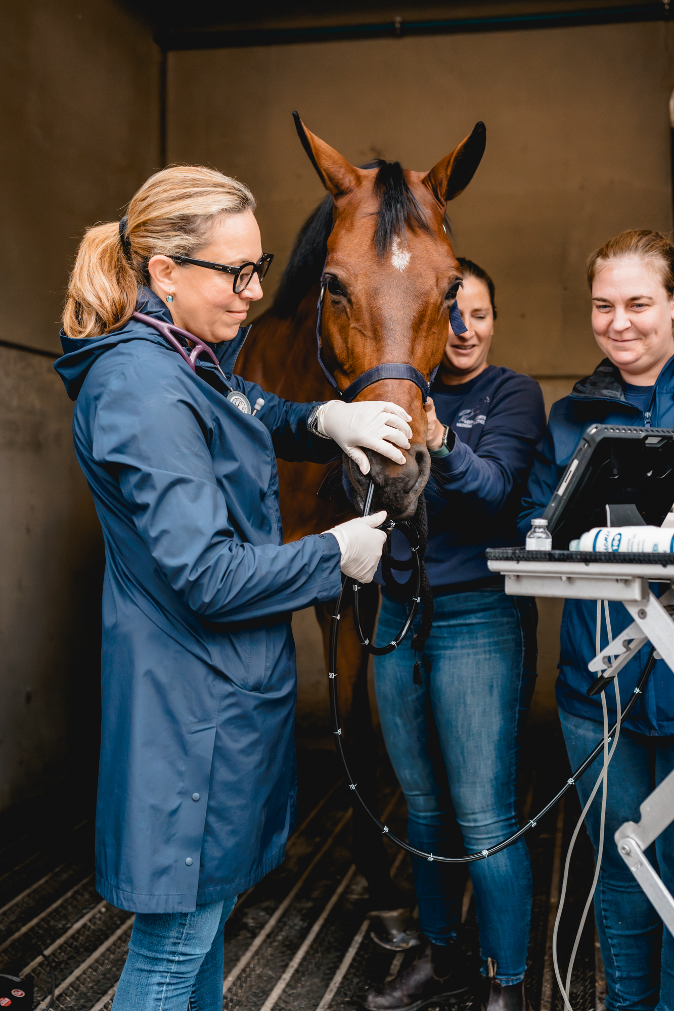
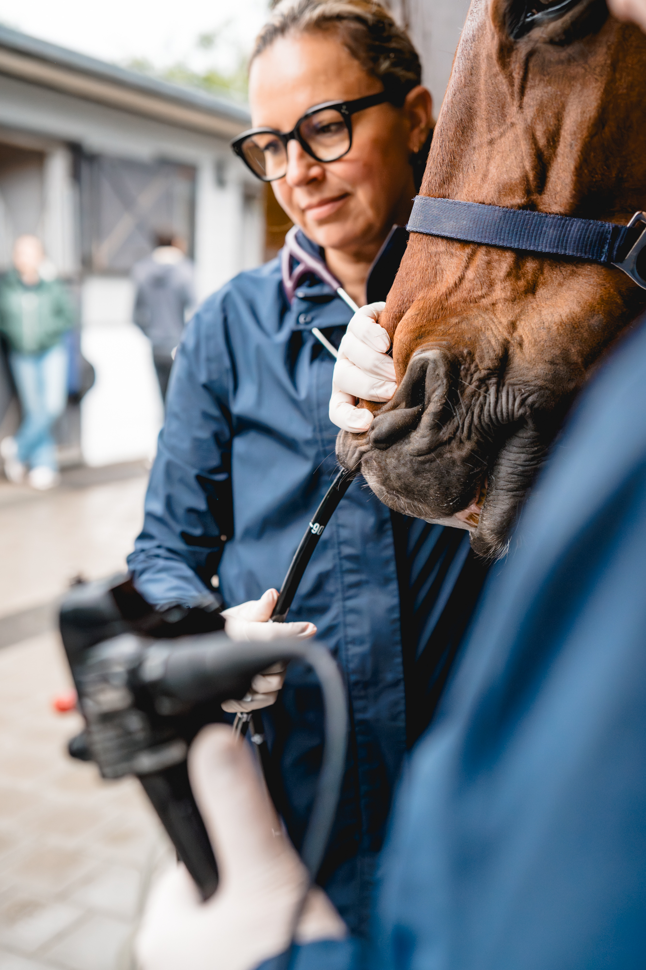
The examination is usually scheduled in the morning. The horse is lightly sedated and a flexible endoscope is gently passed through the nostril and oesophagus into the stomach.
The different regions of the stomach are then carefully examined, allowing any abnormalities to be identified and documented.
Intestinal Biopsy
What is an Intestinal Biopsy?
When intestinal disease is suspected, a small sample of intestinal mucosa can be collected and submitted to a specialised laboratory for microscopic examination.
Histopathological analysis can provide valuable information in cases of suspected inflammation, infiltration, infection or other intestinal abnormalities, and can contribute to establishing a more precise diagnosis.
How is an intestinal biopsy performed?
In selected cases, intestinal tissue samples can be obtained from a sedated, standing horse.
Depending on the area that needs to be investigated, samples may be collected from the proximal small intestine during gastroscopy or from the rectal mucosa.
A specialised biopsy instrument is used to collect a very small tissue sample for laboratory analysis. The procedure does not usually require sutures.
Abdominal ultrasonography
What is an abdominal ultrasonography?
Abdominal ultrasonography is a non-invasive imaging technique that allows us to examine many of the structures within the horse’s abdomen.
It can provide valuable information about portions of the intestinal tract, liver, spleen, kidneys and stomach, including structures that cannot be fully assessed through a transrectal examination.
Ultrasonography can help identify changes in organ appearance, intestinal wall thickness, motility and abdominal fluid, and is therefore an important part of the investigation of many digestive and abdominal conditions.
How is this examination performed?
Abdominal ultrasonography is performed with the horse standing and sedation is often unnecessary.
Depending on the horse and the area being examined, small areas of hair may need to be clipped. The skin is prepared with an appropriate contact medium to ensure optimal ultrasound image quality.
Absorption Test
What is it?
In horses with chronic or unexplained weight loss, it may be necessary to assess whether nutrients are being adequately absorbed through the intestinal tract.
An oral glucose absorption test evaluates the horse’s ability to absorb a measured amount of glucose administered directly into the stomach. Changes in blood glucose levels over time provide information about intestinal absorptive function.
How is this test performed?
The horse is fasted before the examination. An initial blood sample is collected to establish a baseline blood glucose concentration.
A measured amount of glucose solution is then administered into the stomach through a nasogastric tube. Blood samples are collected at regular intervals over the following hours.
The resulting glucose curve is analysed to assess whether intestinal absorption appears normal, delayed or reduced.
Abdominal Paracentesis
What’s abdominal paracentesis?
The abdominal organs are surrounded by a small amount of fluid known as peritoneal fluid.
Changes in the appearance, composition or cellular content of this fluid can provide important information when abdominal disease is suspected. Collecting and analysing a sample can therefore contribute to the investigation of conditions involving inflammation, infection, intestinal disease or other abdominal abnormalities.
How is this examination performed?
The procedure is performed with the horse standing and, when required, lightly sedated.
A small area underneath the abdomen is clipped and carefully disinfected. A sterile needle or cannula is then introduced into the abdominal cavity to collect a small sample of peritoneal fluid.
The sample can be evaluated and, when indicated, submitted for further laboratory analysis.
Our clinical services
Is something affecting your horse's health, well-being or performance?
Looking for answers about your horse's health, well-being or performance?
Our team combines clinical expertise with advances diagnostic tools to support your horse’s health and well-being at every stage.